
📌 Introduction to the History of Optical Microscopes
The optical microscope is among the major inventions that have revolutionized our understanding of the living world and matter. Introduced centuries ago, this instrument has allowed researchers and enthusiasts to observe, for the first time, details invisible to the naked eye. Its impact on science extends from biology to medicine, as well as physics and chemistry. The history of the optical microscope is part of a constant quest to improve human perception, generating essential discoveries and advancements in many fields.
Today, before revealing its remarkable evolution, let's explore the origin and fundamental role of the optical microscope in the scientific revolution. Understanding the beginnings of this device enlightens us on how it has transformed observation, research, and scientific pedagogy throughout the centuries. From its modest origins to sophisticated digital models, the optical microscope has undergone an evolution guided by human ingenuity and the need to know ever more about the living and non-living.
Let's delve into a fascinating history of ingenuity, revolutionary discoveries, rivalries, and international collaborations, showing that the tiny world discovered by the optical microscope has had vast repercussions on our understanding of nature and on modern society.
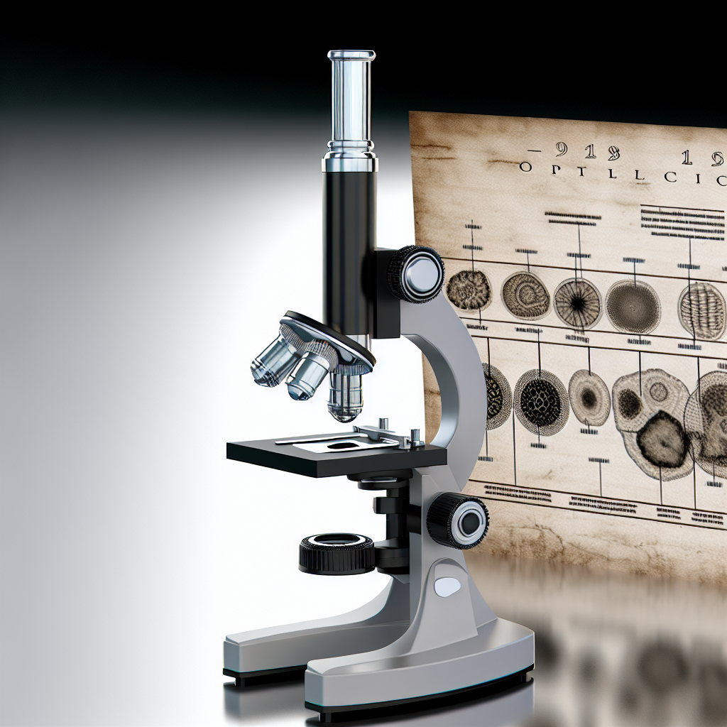
📌 The First Microscopes: A Historical Perspective
The development of the optical microscope began between the end of the 16th century and the beginning of the 17th century, marked by ingenious experiments with optical lenses. The first rudimentary microscopes appeared thanks to glass artisans, who discovered that two lenses aligned one behind the other magnified tiny objects. Thus began the first explorations of the microscopic world, redefining the limits of what is observable.
At that time, the nature of lenses and their properties were relatively poorly understood, but the thirst for discovery led scientists and inventors to perfect these instruments. Several figures quickly emerged, including Zacharias Janssen, often mentioned as the inventor of the first optical microscope, as well as Galileo Galilei, who made significant technical improvements.
These pioneers laid the foundations for a long line of innovators, initiating the metamorphosis of what was once just a curious optical toy into an indispensable scientific tool. Their first prototypes, though simple compared to modern devices, already opened the doors to an invisible and fascinating world.
📌 The Influence of Zacharias Janssen and Galileo Galilei
Zacharias Janssen, a Dutch artisan, is traditionally credited with the invention of the microscope around 1590, although his exact involvement is a subject of debate among historians. According to tradition, Janssen, with the help of his father Hans, had the idea of aligning two lenses to obtain unprecedented magnification, thereby giving birth to the first compound microscope. However, the documentation from that period remains fragmentary.
In parallel, Galileo Galilei, an Italian scholar primarily known for his astronomical discoveries, also took an interest in optics. Around 1609, he designed an instrument called "occhiolino" (little eye), which utilized improved convex lenses. Galileo precisely described his devices and carried out some of the earliest observations of microscopic life, such as patterns on insect shells and botanical details.
Galileo's role is fundamental because he applied scientific rigor to his experiments, seeking to perfect image quality and correctly interpret discoveries. His work helped optimize lens alignment and improve the design of microscopes available on the market at the time. Thanks to his notoriety, he motivated other scholars to explore the infinitely small, thus launching the first wave of research into the microscopic world.
This era therefore marks the beginnings of optical microscopy, where the spirit of innovation and scientific curiosity paved the way for increasingly advanced techniques, building the foundations of modern microscopy.
📌 Advancements in the 18th Century
During the 18th century, the optical microscope underwent major developments, both in its technical design and in the precision of its lenses and the expansion of its scientific uses. The era was marked by the birth of learned societies and international correspondence, accelerating the dissemination of innovations in the scientific world. The growing needs of biology, anatomy, and entomology stimulated the refinement of this valuable tool.
Many inventors improved optical resolution, robustness, and ease of use, making microscopes more accessible to scientists and students across different disciplines. The quality of the images obtained also improved, thanks to the development of new optical formulas and the use of more performant materials for lens manufacturing.
A notable evolution of this period was the gradual abandonment of the simple microscope in favor of the compound microscope, designed to provide significantly higher magnification power and clarity. The instrument, until then reserved for a minority of scholars or passionate aristocrats, gradually became more popular among educational institutions and nascent laboratories.
The 18th century was thus a time of intense scientific emulation, at the crossroads of technical progress and theoretical innovations, where optical microscopy asserted its key role in the exploration of the invisible.
📌 The Compound Microscope and its Improvements
The transition from the simple microscope to the compound microscope marked a decisive turning point in the history of microscopy. The compound microscope is distinguished by the combination of several lenses – an objective close to the object to be observed and an eyepiece near the user's eye. This configuration, by multiplying the initial magnification, offered images of unparalleled sharpness and detail for the era.
Artisan opticians like John Cuff and Benjamin Martin in England played a pivotal role: they introduced ergonomic designs, stabilized mounts, and perfected the focusing mechanism. Their work resulted in more robust microscopes, equipped with stands allowing prolonged observations without excessive fatigue. The materials used evolved, moving from wood to metal alloys resistant to wear and mechanical shock.
- Introduction of iris diaphragms, allowing control of incident light intensity and adjustment of image contrast.
- Appearance of plane or concave mirrors, facilitating light reflection onto the observed sample.
- Development of fine focusing systems, essential for obtaining sharp images.
All these innovations contributed to democratizing the use of the compound microscope within scientific circles. New models were marketed in universities and learned societies, which fostered the multiplication of anatomical, botanical, or zoological observations.
The compound microscopes of the 18th century thus prepared the ground for the explosion of scientific discoveries that would follow, by making optical microscopy more reliable, powerful, and accessible.
📌 The Golden Age of Microscopes in the 19th Century
The 19th century truly represents the golden age of the optical microscope, a period during which its role in major scientific discoveries reached its peak. This instrument became a fundamental tool in the race to understand biological, medical, and chemical phenomena. Progress in glass manufacturing, combined with advances in physical optics, led to the creation of achromatic and apochromatic lenses, correcting chromatic aberrations responsible for image distortion.
It was during this time that microscopes became powerful enough to reveal the complexity of living cells, fostering the birth of new disciplines. Cellular structure, bacteria, protozoa, and unknown tissues were identified thanks to unprecedented observations. The work of Robert Brown (discovery of the cell nucleus), Matthias Schleiden and Theodor Schwann (cell theory), and Rudolf Virchow (omnis cellula e cellula) revolutionized biology thanks to the optical microscope.
Learned societies and renowned laboratories acquired increasingly sophisticated models. Major firms such as Carl Zeiss, Ernst Leitz, and Nachet established themselves in the market, constantly innovating to meet the growing demand from researchers and doctors.
The tool, once reserved for a few initiates, became an international reference in scientific research. It shaped the destiny of modern biology and medicine by structuring observation protocols and codifying microscopic investigation.
📌 The role of Charles Darwin and other naturalists
At the heart of the 19th century, the explosion of naturalist research is inseparable from the advances made thanks to the optical microscope. Charles Darwin, famous for his theory of evolution, regularly used this invaluable instrument in his extensive travels aboard the Beagle or in his subsequent studies. Darwin observed, dissected, and analyzed samples from all continents, discovering a fascinating diversity of structures and life forms, from microorganisms to mollusks.
The microscope allowed Darwin to gather meticulous evidence on the variability and adaptations of species. He described cellular arrangements, morphologies of embryos or tissues, which guided his thinking on evolution. Following in his footsteps, many other naturalists and biologists – such as Louis Pasteur, Ferdinand Cohn, Anton de Bary, and Ernst Haeckel – utilized microscopy to study microbes, fungi, plants, and animals.
The research conducted by these scholars led to major discoveries:
- Isolation and identification of the first pathogenic bacteria.
- Study of plant and fungal reproduction at the microscopic scale.
- Understanding of cell structure and organization of living tissues.
The optical microscope thus became an indispensable vector for advancing knowledge, providing a new generation of researchers with the means of investigation they had always dreamed of.
Thanks to these advances, biology, medicine, and chemistry emancipated themselves, laying the foundations for a modern, rigorous, and empirical science.
📌 The 20th Century Scientific Revolution
The 20th century revolutionized optical microscopy, not only through the multiplication of technological innovations but also through the integration of converging disciplines such as quantum physics, engineering, and electronics. From then on, research focused on increasingly fine resolutions, capable of revealing sub-cellular structures previously out of reach.
The constant improvement of optics and light sources made it possible to observe ultra-precise details, facilitated by monochromators, fluorescence filters, and discharge lamps or LEDs. Simultaneously, the advent of the electron microscope in the 1930s further pushed the observation capabilities of the microscopic world, reaching magnifications of over 1,000,000 times in some cases.
Yet, the optical microscope retained all its relevance, integrating into hybrid, automated, and versatile devices, serving medical imaging, advanced cell biology, and industrial control.
This was also the century when microscopy became widely adopted in school and university education, training generations of scientists. The microscope became a central tool for tissue analysis, cell cultures, medical pathology, and material analysis.
The instrument then came in a variety of specialized versions, each addressing specific challenges in biology, biomedicine, or the pharmaceutical industry.
📌 The Emergence of New Types of Microscopes
The most significant innovation of the 20th century was undoubtedly the emergence of microscopes utilizing new physical principles to achieve unparalleled resolutions. In addition to the refinement of the classic optical microscope, several variants emerged:
- Transmission Electron Microscope (TEM): Replaces light with an electron beam, allowing observation of the internal structure of cells and macromolecules at a nanometric scale.
- Scanning Electron Microscope (SEM): Scans the surface of samples to generate three-dimensional images with remarkable detail.
- Atomic Force Microscope (AFM): Uses interactions between an ultrafine tip and the surface of a sample to map atomic reliefs.
- Fluorescence and Phase Contrast Microscope: Exploiting the properties of light on biological components, it reveals the localization or activity of specific molecules after labeling.
While these tools push the boundaries of resolution, their complementarity with the traditional optical microscope is evident: many modern laboratories combine several approaches to explore both the global structure and the molecular detail of samples.
To discover a comprehensive overview of recommended current models and the advantages of each type, consult our dedicated page on advanced optical microscopes.
This instrumental abundance considerably broadened the range of tools available to researchers, who can now choose the most appropriate technique for each scientific question.
📌 Modern Microscopes and 21st Century Innovations
The last two decades have witnessed a new wave of innovations in optical microscopy, driven by the rapid pace of technological progress, miniaturization, and digital convergence. Modern microscopes have become true analytical stations, combining precision, versatility, and ease of use.
Among the significant new features are digital microscopes, capable of capturing high-resolution images, making video recordings, and instantly sharing observations via connected networks. These instruments are equipped with powerful software for image analysis, 3D reconstruction, and quantification of specific features.
The rise of global collaborations and the trend towards multidisciplinarity also benefit microscopy, a true pivot for exchanges between biologists, engineers, and computer scientists. Ergonomics have significantly improved, with touch interfaces, motorized stages, and automated systems allowing for high-throughput analyses.
The scope of the optical microscope continues to expand: it is now indispensable in advanced laboratories as well as in education, medicine, the agri-food industry, research on innovative materials, and environmental studies.
📌 The Impact of Digitization and Artificial Intelligence
The integration of digitization and artificial intelligence (AI) is radically transforming contemporary microscopy. Intelligent algorithms automate many tasks, such as pattern recognition, cell classification, anomaly detection, and image reconstruction from massive data.
Digitization allows millions of sample images to be stored and accessed in remotely viewable databases — fostering autonomous learning and comparison between research centers worldwide. Collaborative platforms accelerate the sharing of images and results, contributing to more accessible and faster science.
A key advance lies in automated image analysis: for example, thanks to AI, screening for cancer cells in tissues, identifying pathogens, or automatically measuring anatomical structures become more reliable and faster. Microscopes connected to computer networks actively participate in telemedicine, remote diagnosis, and interactive training for students or young researchers.
Finally, augmented reality and virtual interfaces open up unprecedented educational prospects, making microscopy more attractive to new generations.
📌 Contributions of Microscopes to Modern Biology
The optical microscope remains the cornerstone of major discoveries in modern biology. Thanks to it, cell theory was formalized in the 19th century; this advance laid the foundations for genetics, pathology, and physiology. Direct observation of cells and their organelles—mitochondria, chloroplasts, nuclei, etc.—has made it possible to understand their functioning, their life cycle, and their involvement in health or disease.
Today, biologists use microscopes to study the mechanisms of embryonic development, cellular aging, tissue differentiation, and immunity. Major discoveries, such as the structure of DNA, cell division (mitosis, meiosis), and cell migration, have been made possible thanks to microscopy.
Advances in molecular biology, such as the use of labeled antibodies or fluorescent dyes, also depend on the analytical power of modern microscopes. In medical research, the diagnosis of cancers, infections, or genetic diseases still relies on microscopic examination of tissues, cells, or biological fluids.
The contributions of the optical microscope are not limited to fundamental research. It plays a central role in the development of new drugs, the study of infectious agents, the characterization of microorganisms in the food industry, and water quality monitoring. Its versatility ensures its sustainable future, in close collaboration with advanced techniques such as cytometry and confocal microscopy.
📌 Advanced Techniques and Current Applications
Constant technological progress has given rise to advanced optical microscopy techniques, multiplying observation capabilities tenfold. Among the most recognized are fluorescence microscopy, super-resolution microscopy, confocal microscopy, and multiphoton microscopy.
Fluorescence microscopy allows the localization and quantification of specific molecules in cells by exploiting the light emitted by fluorophores. Coupled with antibodies or genetic probes, it can be used to study the distribution of proteins, RNA, or other biomolecules in real time.
Super-resolution microscopy overcomes the limitations imposed by light diffraction, allowing the observation of structures at the nanometric scale, such as cytoskeletal protein networks or macromolecular complexes. These methodologies—PALM, STED, SIM, among others—open new horizons in the study of cellular mechanisms.
Current applications extend to hospital research, genetics, pathology, biotechnology, and the pharmaceutical industry. Automated laboratories use robotic microscopes for high-throughput analysis of thousands of samples simultaneously, accelerating drug development and the validation of innovative treatments.
The versatility of these techniques ensures that optical microscopy maintains an essential place in medical research strategies, environmental monitoring, and the improvement of industrial practices.
To discover our selection of the best tools on the market, read our comparative article "Top 5 Recommended Optical Microscopes This Year" and choose the one that suits your scientific or educational needs.
📌 Optical Microscopy and Scientific Education
The use of the optical microscope in scientific education has profoundly influenced how students have learned biology, physics, and chemistry since the 19th century. From primary school, the discovery of the microscopic world sparks passion and wonder. Teachers use microscopes to explain the cell, the diversity of tissues, the structure of leaves, hair, and insects.
In middle and high school, microscopy becomes a vehicle for structured and concrete learning—whether it's making observations, preparing slides, or understanding the anatomy of living organisms. Practical experiments facilitate memorization and understanding of complex concepts such as cellular reproduction, microorganisms, and genetics.
At university, the instrument is inseparable from fundamental and applied research. Students in biology, medicine, geology, and materials science receive in-depth training in optical microscopy, which is essential for their scientific careers. Tissue analysis, cytology, pathology, ecology, and materials study rely on brief manipulations with the microscope.
With the advent of digital and portable models, access to microscopy is becoming even more widespread. Schools now have robust, connected tools that allow live sharing of observations on screen—an essential factor in attractiveness and equal opportunities in scientific learning.
📌 Economic and Industrial Impact of Optical Microscopes
Beyond research and education, the optical microscope holds a growing place in modern industry. Its influence extends to pharmaceutical production, material quality control, microelectronics, agro-food, and environmental applications.
In the pharmaceutical sector, it allows for checking the purity of active ingredients, detecting impurities, and validating the manufacturing quality of medicines. In the semiconductor industry, it is essential for inspecting the quality of integrated circuits and the regularity of nanometric structures.
The agro-food and cosmetic industries use microscopy to identify contaminants, control product texture, and certify batch compliance. Environmental control laboratories use it to monitor the presence of microorganisms or harmful particles in water, air, and soil.
The instrument thus participates in essential industrial processes, contributing to business competitiveness, consumer safety, and product traceability. Its integration into production chains via automation and robotics promotes productivity and quality, ensuring a major economic impact.
Finally, the growing demand for portable microscopes and connected tools accelerates innovation, creating a dynamic and growing market in the scientific technology sector.
📌 Challenges and Future of Optical Microscopy

Within 14 days - Unconditional

Products tested and approved

Support a local business

Guarantee of a Smooth Transaction


Research